In any ophthalmological examination it is not considered complete without indirect ophthalmological examination. Usually after slit lamp examination in routine examination ,your doctors asks for you pupils to be dilated. For purpose of dilating usually some drops are put in your eyes. These drops are Tropicamide or Homatropine bromide. Dilated examination has mainly two purpose. first to know the exact glass power and secondly to examine the back of the eye or retina of the eye.
Retinal examination include the central and peripheral examination. Central retinal examination can divulge the details of central part where macular diseases , glaucoma, ARMD, Macular hole, Diabetic retinopathy, retinal vascular occlusion etc. can be diagnosed. Peripheral retinal examination is also of utmost important to rule out retinal lattice, atrophic retinal holes, retinal tears, intermediate uveitis or vascular occlusion, sometimes early diabetic retinopathy.
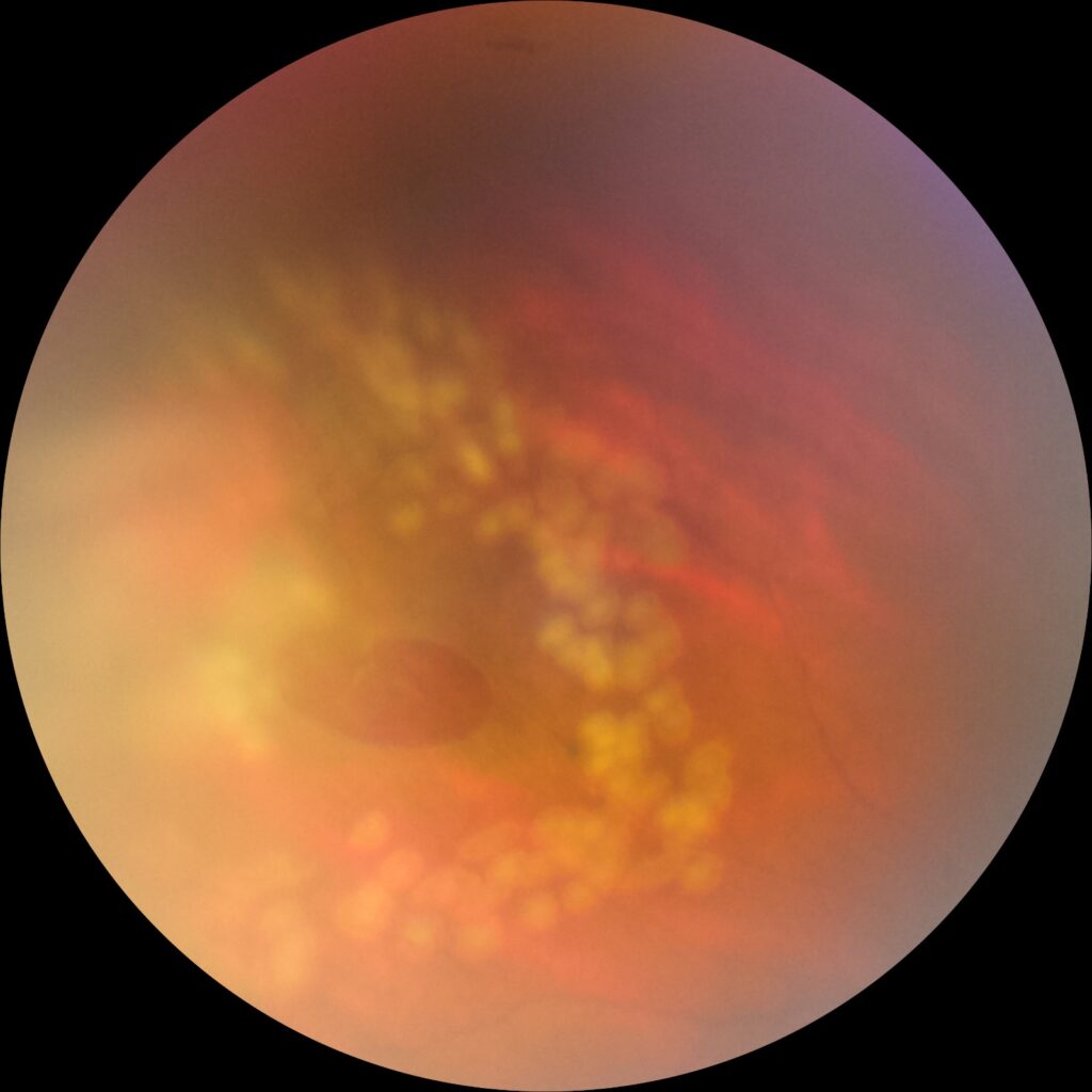
Lattice degeneration – theses are degeneration of peripheral retina. In these areas the retina is thinned out and weak, over these areas there is adhesions of peripheral vitreous and in case of traction during the event of posterior vitreous detachment there are great risk of tear formation around theses lesions. Theses tears can result in detachment of retina. During the event of traction the patient can complain of floaters and flashes. So it is important for general masses to know the importance of flashes and floaters. Any type of FLASHES OF LIGHT and sudden appearance of HUNDREDS OR THOUSANDS OF BLACK SPOTS in field of vision should be reported to any ophthalmologist in nearest available location.
Any type of FLASHES OF LIGHT and sudden appearance of HUNDREDS OR THOUSANDS OF BLACK SPOTS in field of vision should be reported to any ophthalmologist in nearest available location.
If diagnosed at time and properly lasered the catastrophic event of retinal detachment can be prevented. A barrage is made surrounding the lattice with help of argon green laser. Theses laser spots strengthen the surrounding retina and can reduce the further chances of retinal detachment in up to 95% of cases.

lattice degeneration of retina
Here is a picture of peripheral retina showing the lattice degeneration. Usually these are spread circumferentially but there are also radial lattice degeneration along the retinal vessels.